
Both methods measure soft tissue stiffness, but they are not interchangeable, so pick one and track trends within it. Shear wave elastography uses an ultrasound machine to image stiffness through tissue depth and is the research gold standard. Myotonometry uses a handheld probe at the skin surface and reports stiffness in N/m within seconds. A 2024 review in SN Comprehensive Clinical Medicine found the agreement between the two ranges from moderate to strong depending on the muscle, examiner, and population, with real discrepancies in some cases. A 2025 validation study added that subcutaneous fat measurably affects the handheld reading, so the choice usually comes down to whether you need imaging context.
What is the core difference between the two methods?
The measurement physics are different, which is why the units differ.
Shear wave elastography (SWE) is an ultrasound technique. A focused acoustic pulse generates a transverse shear wave inside the tissue, and the speed of that wave is converted into a Young's modulus value reported in kilopascals (kPa). The output is a color-coded stiffness map overlaid on the B-mode ultrasound image, so the clinician can see both anatomy and stiffness in the same frame.
Myotonometry is a mechanical impulse technique. A spring-loaded probe (such as the MyotonPRO or comparable handheld devices) delivers a brief tap to the skin and records the resulting damped oscillation. From that oscillation it reports stiffness in N/m, frequency in Hz, decrement, relaxation time, and creep. There is no image, only a number, but the number is reproducible within seconds.
Do the two methods agree?
Yes, with caveats. A 2021 study in Diagnostics found that shear wave elastography and the MyotonPRO produced correlated stiffness readings across major muscles, supporting handheld myotonometry as a portable alternative to ultrasound elastography. The absolute values cannot be compared directly because the units differ (kPa for SWE versus N/m for myotonometry), but trends and within-patient changes track together.
For routine clinical comparison over a care plan, what matters is direction and magnitude of change, not absolute value. Both methods deliver that. For research comparing across populations, SWE values converted to Young's modulus are more standardized.
How reliable is each method?
Both have strong reliability data in recent literature.
A 2024 systematic review in Medicina of 48 studies across 31 muscle groups found good-to-excellent intra-rater and inter-rater reliability for the MyotonPRO. A 2024 study in Frontiers in Sports and Active Living reported ICC values of 0.74 to 0.99 across most lower-extremity muscles in athletes. SWE has comparable reliability when performed by trained operators with standardized probe placement.
Both modalities are sensitive to patient position, time of day, and muscle activation state. A 2026 lumbar erector spinae myotonometry study found that posture, spinal level, gender, and muscle activation state all significantly affected the reading. The same is true for SWE. A written protocol is required for either tool to produce comparable values across visits.
Survey data: In a 2026 survey of 455 patients who stopped chiropractic care, 58% cited perception-based reasons: 36% felt no progress, and 22% felt better and stopped. Neither group was told their stiffness was still elevated.
Side by side: which fits which workflow?
| Factor | Myotonometry (e.g., MyotonPRO) | Shear wave elastography |
|---|---|---|
| Cost | Roughly $5,000 to $15,000 for the device | Roughly $40,000 to $200,000 for the ultrasound system with SWE module |
| Training | Self-taught with a written protocol, ~2 hours to proficiency | Sonography training plus elastography certification, ~weeks to months |
| Time per point | 30 to 60 seconds | 2 to 5 minutes including probe positioning |
| Depth of measurement | Superficial, ~1 to 2 cm below probe | Full muscle thickness, can reach deep structures |
| Output | Numerical value (N/m, Hz, decrement) | Color stiffness map overlaid on B-mode image |
| Portability | Handheld, battery-powered | Cart-based or laptop-based with probe |
| Best fit | Chairside re-exams, longitudinal tracking, patient-facing reports | Diagnostic imaging, research, deep muscles, complex cases |
Which one should a chiropractor or PT choose?
For chairside use in private practice, myotonometry is usually the better fit. The workflow looks like this: take a baseline reading at intake, repeat at the visit-6 or visit-12 re-exam, and show the patient the comparison. The reading takes under a minute, the device sits on the desk, and the patient leaves with a number they can talk about.
SWE makes sense when imaging is already part of the practice (sports medicine, MSK ultrasound clinics, hospital-affiliated rehab), when deep muscles need to be measured (psoas, deep lumbar multifidus), or when the diagnostic question requires resolving structure as well as stiffness. A 2024 study showed that myotonometry combined with extended field-of-view ultrasound allowed reliable quantification of patellar tendon stiffness and length at rest and during load, which is an example where the two methods complement each other rather than substitute.
What are the practical limitations of each?
Myotonometry limitations: superficial measurement only, single-point sampling (so you measure the muscle at one spot, not a region), and no imaging context to confirm landmark. Skin thickness, body composition, and probe pressure can affect the reading if the protocol is not standardized.
SWE limitations: cost, training time, dependence on operator skill, probe pressure sensitivity, and lower throughput. SWE values can also be affected by tissue anisotropy (fiber orientation), so probe alignment to muscle fiber direction is part of the protocol.
What about other handheld options?
Algometers measure pressure pain threshold, not stiffness directly. Durometers measure surface hardness and are sometimes adapted for soft tissue work but lack the validation base of myotonometry. A 2025 study in PeerJ reported clinical reliability and responsiveness for tissue hardness meters in musculoskeletal contexts, but the literature is much thinner than for myotonometry or SWE. For a defensible objective measure, the two methods with substantial peer-reviewed support are myotonometry and shear wave elastography.
Frequently Asked Questions
How does myotonometry compare to shear wave elastography?
Both measure soft tissue stiffness and produce correlated readings. SWE uses ultrasound to image stiffness through depth; myotonometry uses a handheld probe at the skin surface and reports N/m within seconds. Myotonometry is portable and cheap; SWE adds imaging context.
Do the two methods produce the same numbers?
No, the units differ (kPa for SWE versus N/m for myotonometry), but trends and within-patient changes track together. A 2021 Diagnostics study confirmed correlated readings across major muscles.
Which is better for a chiropractic or PT practice?
Myotonometry usually fits private practice better because it is fast, portable, and patient-facing. SWE makes sense when imaging is already part of the workflow or when deep muscles need to be assessed.
How long does each method take per measurement?
Myotonometry takes 30 to 60 seconds per point. SWE takes 2 to 5 minutes per measurement including probe positioning and image acquisition.
Can a handheld myotonometer reach deep muscles?
Not directly. Myotonometry measures the superficial tissue volume below the probe, typically 1 to 2 cm. For deep muscles like psoas or deep lumbar multifidus, SWE has the advantage.
Are both methods reliable across raters?
Yes when the protocol is standardized. A 2024 Medicina systematic review of 48 myotonometry studies found good-to-excellent intra-rater and inter-rater reliability. SWE shows comparable reliability with trained operators.
What about portable ultrasound elastography?
Portable SWE devices are emerging but still require sonography training and standardized probe alignment. For a chairside workflow with under-a-minute measurement time and no imaging interpretation, handheld myotonometry remains the lower-friction option.
Does body fat affect myotonometry readings?
Yes. A 2025 study comparing the MyotonPRO to shear wave elastography found that subcutaneous fat thickness had a significant moderate-to-strong effect on handheld stiffness readings, and the two methods could not be used interchangeably for the rectus femoris. The practical takeaway is to track trends within one device rather than comparing absolute numbers across methods or across patients with very different tissue composition.
How well do myotonometry and shear wave elastography agree?
A 2024 review in SN Comprehensive Clinical Medicine reported that agreement between the two methods ranges from moderate to strong depending on the muscle, examiner, and study population, with meaningful discrepancies in some settings. The two capture related but distinct properties, so a reading from one should not be treated as a substitute for the other.
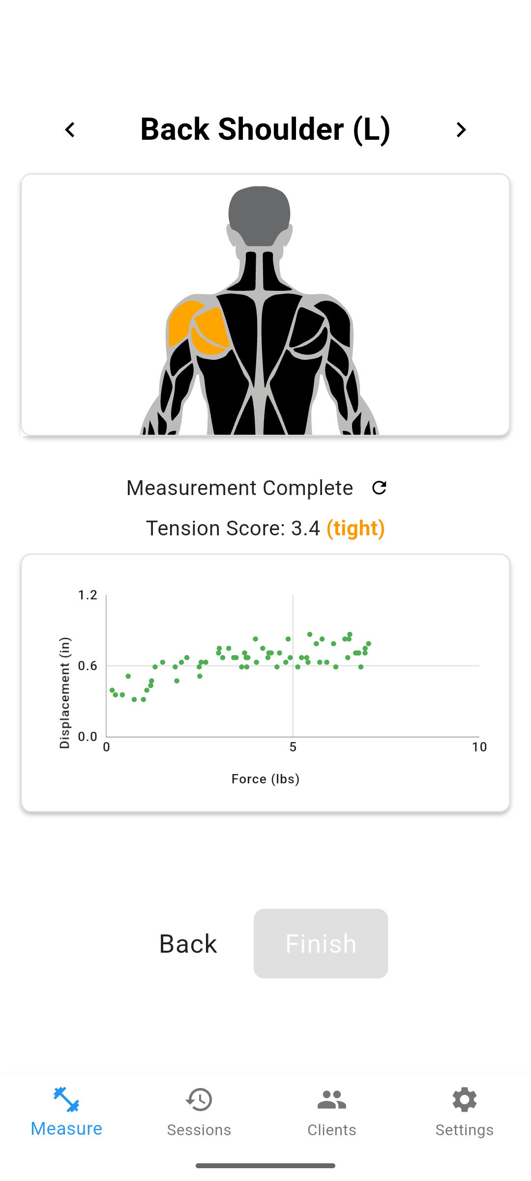
One approach is to add a second channel of objective data alongside subjective pain reports. Options include soft tissue stiffness measurement (such as MuscleMap), range-of-motion testing, and posture analysis. Each gives you something concrete to show the patient rather than asking them to take your word for it.