
Measure the tibialis anterior with a handheld myotonometer or shear wave elastography, at a standardized position with the ankle relaxed. Place the patient supine or seated, find the muscle belly on the front-outer shin, and press the probe perpendicular to the skin. The single most important rule is consistency: same position, same point, same ankle angle every visit, so readings are comparable.
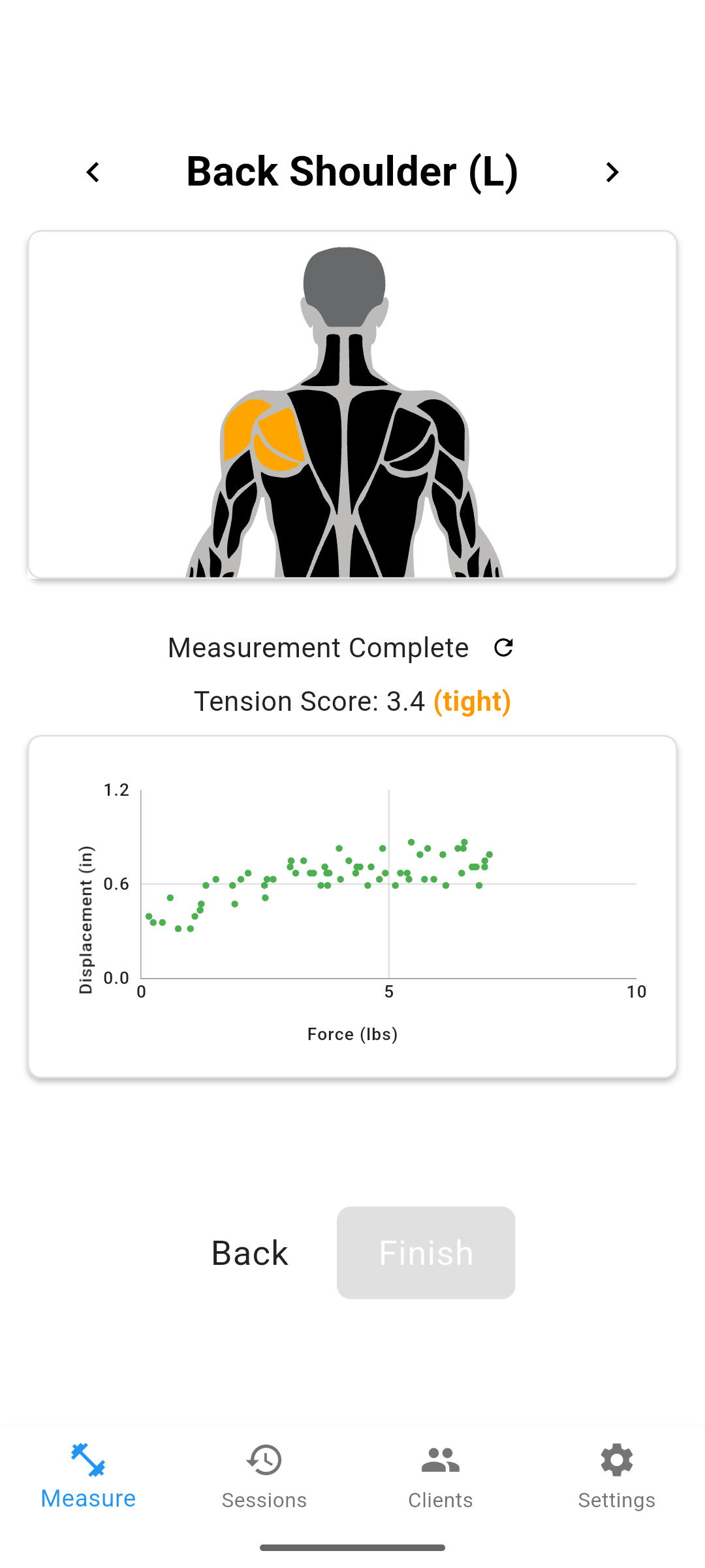
Where Is the Tibialis Anterior Measurement Point?
On the front-outer shin, about a third of the way down from the knee, just lateral to the tibia. Have the patient actively dorsiflex the ankle so the muscle belly stands out, palpate for its thickest part, and mark it. Then relax the ankle before taking the reading. Marking the exact spot matters because moving even a centimeter along the muscle can change the value you get, and you want the same landmark at every re-exam.
Myotonometer or Shear Wave Elastography?
Both work, and they agree reasonably well. A quantitative study in Diagnostics measured stiffness of major lower limb muscles, including the tibialis anterior, with both shear wave elastography and a myotonometer, and reported moderate to strong correlations between the two methods at rest and during contraction. For a chairside physical therapy setting, a handheld myotonometer is faster and does not require imaging, while elastography can give more detail on deep versus superficial fibers. A 2025 study found that stiffness of the deeper tibialis anterior fibers related to functional recovery after ankle fracture surgery.
Should the Ankle Be Relaxed or Contracted?
Either is valid, but choose one and keep it fixed. Muscle stiffness reads lower at rest and higher during voluntary dorsiflexion, so mixing states across visits will produce noise that looks like real change. For tracking a patient over time, resting measurements with the ankle neutral and supported are the easiest to reproduce. Age also shifts the baseline: a study using both elastography and a myotonometer found lower tibialis anterior stiffness in older adults than younger adults, which is why each patient should be compared to their own baseline.
| Variable | Standardize it to | Why it matters |
|---|---|---|
| Body position | Supine or seated, same each time | Position changes resting tone |
| Measurement point | Marked muscle belly, front-outer shin | A shift of 1 cm changes the value |
| Ankle state | Relaxed neutral, or fixed contraction | Rest reads lower than contraction |
| Reference | Patient's own baseline | Age and individual variation are large |
Survey data: In a 2026 survey of 455 patients who stopped chiropractic care, 58% cited perception-based reasons: 36% felt no progress, and 22% felt better and stopped. A repeatable objective reading, whether at the tibialis anterior or elsewhere, gives you a concrete number to show a patient who cannot feel the change you are tracking.
Frequently Asked Questions
How do you measure tibialis anterior stiffness?
Place the patient supine or seated with the ankle relaxed in a neutral position, locate the tibialis anterior muscle belly on the outer shin, and take the reading with a handheld myotonometer pressed perpendicular to the skin or with shear wave elastography. Keep the position and probe angle the same every time so readings are comparable.
Where is the tibialis anterior measurement point?
The muscle belly is on the front-outer aspect of the shin, roughly a third of the way down from the knee toward the ankle, just lateral to the tibia. Palpate for the thickest part of the muscle while the patient dorsiflexes, then relax the ankle before measuring.
Is myotonometry reliable for the tibialis anterior?
Yes, for a single trained rater using a standardized position. Studies comparing myotonometry with shear wave elastography on lower limb muscles including the tibialis anterior report moderate to strong correlations between the two methods, both at rest and during contraction.
Should the ankle be relaxed or contracted during measurement?
Pick one and stay consistent. Stiffness is lower at rest and higher during voluntary dorsiflexion, and both states are measurable. For tracking change over time, resting measurements with the ankle in a neutral, supported position are the easiest to reproduce.
Does age affect tibialis anterior stiffness readings?
Yes. Research using both shear wave elastography and a myotonometer found lower tibialis anterior stiffness in older adults than in younger adults, at rest and during contraction. Compare a patient to their own baseline rather than to a single universal number.
One approach is to add a second channel of objective data alongside subjective pain reports. Options include soft tissue stiffness measurement (such as MuscleMap), range-of-motion testing, and posture analysis. Each gives you something concrete to show the patient rather than asking them to take your word for it.