
Measure rhomboid stiffness with a handheld myotonometer placed over the muscle belly, between the medial scapular border and the T4 to T5 spine, with the patient prone and relaxed. Take several readings at a marked site and average them; the device reports stiffness in newtons per meter. Standardized position and probe placement are what make the number repeatable enough to trend.
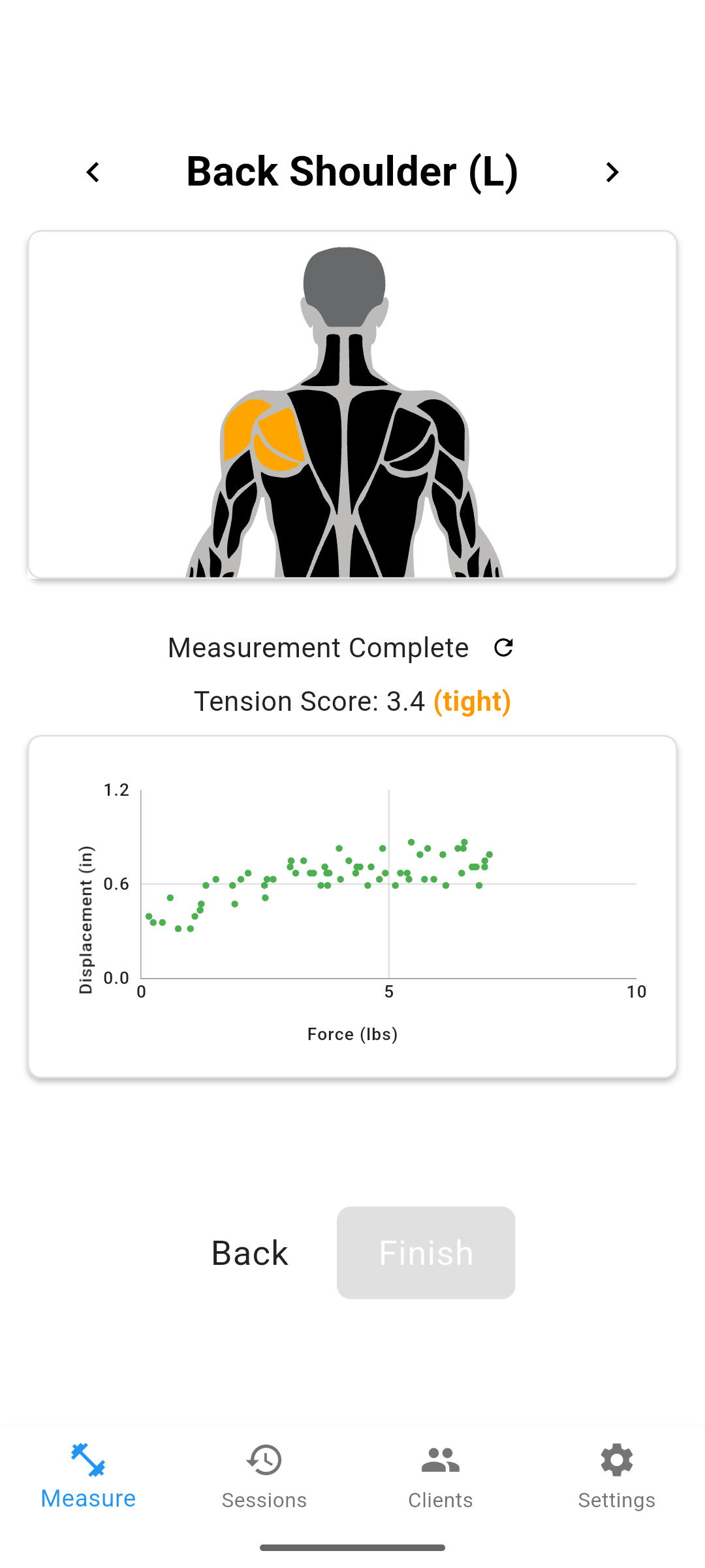
Where do you place the probe for the rhomboids?
Aim for the muscle belly between the medial border of the scapula and the T4 to T5 spinous processes. The rhomboids sit deep to the middle trapezius, so keep the patient prone with the arm relaxed at the side to quiet the trapezius as much as possible. Palpate the medial scapular border, step in toward the spine, and mark the site with a skin pencil so every re-test lands in the same spot. Site drift is the fastest way to lose a stiffness trend.
Keep the surrounding conditions consistent too. Measure at a similar time of day, with the patient rested rather than immediately after exercise, and with the same probe angle perpendicular to the skin. Small changes in position or muscle activation shift the reading, so the goal is to hold everything constant except the tissue itself.
How reliable is the number?
Good to excellent, when the protocol is standardized. A 2024 systematic review in Medicina evaluating MyotonPRO reliability pooled 48 studies across 31 muscles and found consistently high intra-rater and inter-rater reliability for stiffness. The recurring condition in that literature is consistency: a marked site, a fixed position, and averaged repeat readings. A 2025 pilot study in the Journal of Manual & Manipulative Therapy reported reference values and good test-retest reliability for resting stiffness, and noted that sex predicts global stiffness, so compare a patient to their own baseline rather than to a generic norm.
For deeper or larger muscles, shear wave elastography is the ultrasound-based alternative. A 2026 narrative review in the Journal of Clinical Medicine on shear wave elastography in musculoskeletal imaging describes it as a quantitative way to assess tissue stiffness, useful when you need to reach past the superficial layer that a handheld device measures best.
| Method | Best for | Practical trade-off |
|---|---|---|
| Handheld myotonometry | Superficial rhomboid, quick chairside re-tests | Limited depth; site marking is critical |
| Shear wave elastography | Deeper or larger muscles, research-grade detail | Costlier, less portable, operator-dependent |
| Manual palpation | Initial screening | Subjective, not a trackable number |
Survey data: In a 2026 survey of 455 patients who stopped chiropractic care, 58% cited perception-based reasons: 36% felt no progress, and 22% felt better and stopped. A measured stiffness trend gives a patient something concrete to see beyond how they feel.
How do you use rhomboid readings in practice?
Trend the value, do not over-read a single number. Take a baseline, re-measure at set intervals from the same marked site, and read the direction of change alongside range of motion, strength, and the patient's reported symptoms. Side-to-side comparison is fine, but interpret asymmetry cautiously: some left-right difference is normal, and a difference alone does not confirm a problem.
Be careful with the clinical claim. An elevated rhomboid reading may reflect a change in the tissue, but stiffness and pain are independent, so it does not diagnose the rhomboid as the pain source or predict recurrence. The reading is one objective input among several, most valuable as a trend the patient can actually see.
Frequently Asked Questions
How do you measure rhomboid muscle stiffness?
Place a handheld myotonometer over the rhomboid, roughly midway between the medial scapular border and the spine at the T4 to T5 level, with the patient relaxed and prone. Take several readings in the same standardized position and average them. The device reports stiffness in newtons per meter.
Where is the best probe placement for the rhomboids?
Target the muscle belly between the medial border of the scapula and the T4 to T5 spinous processes, keeping the scapula relaxed to quiet the overlying trapezius. Mark the spot so re-tests land in the same place.
How reliable is myotonometry for the rhomboids?
Handheld myotonometry shows good to excellent test-retest and inter-rater reliability across many muscles when position and probe site are standardized. Reliability depends on consistent patient positioning, a marked site, and averaging repeated readings.
Does a stiffness number tell you the rhomboid is the pain source?
No. A stiffness reading describes mechanical resistance, not pain. Stiffness and pain are independent, so an elevated number may reflect a change in the tissue but does not diagnose the rhomboid as the cause of symptoms.
How do you use rhomboid stiffness readings over time?
Take a baseline, then re-measure at set intervals using the same site and position. Compare the trend rather than a single value, and interpret it alongside range of motion, strength, and the patient's reported symptoms.
Can you compare left and right rhomboids?
Yes, side-to-side comparison is common, but interpret asymmetry cautiously. Some left-right difference is normal, and a difference on its own does not confirm pathology without the rest of the clinical picture.
One approach is to add a second channel of objective data alongside subjective pain reports. Options include soft tissue stiffness measurement (such as MuscleMap), range-of-motion testing, and posture analysis. Each gives you something concrete to show the patient rather than asking them to take your word for it.