
Take the reading at a marked point over a relaxed muscle belly, with the limb supported at a fixed joint angle, and compare each result to that patient's own baseline. The setup matters more than the device. And a stiffness number is not a spasticity score, which is the mistake most worth avoiding here.
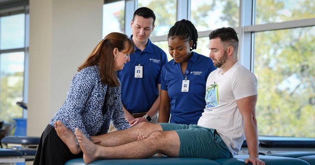
Where do you place the probe, and how do you keep it repeatable?
Pick superficial muscles with a clear belly, mark the site, and lock the joint angle. Published post-stroke work has concentrated on the tibialis anterior, medial gastrocnemius, biceps brachii, and triceps brachii, because all four are easy to find and easy to return to. Mark the point with a skin pencil, note the joint angle and the support surface, and record both alongside the number.
Joint angle is the variable that quietly ruins trends in this population. A spastic limb that rests in a different amount of plantarflexion or elbow flexion from one week to the next will produce a different reading with no change in the tissue. Support the limb the same way each time, let the patient settle for a minute before measuring, and take several readings to average.
Does a stiffness reading measure spasticity?
No, and conflating the two will get you into trouble. Spasticity is a velocity dependent response to passive stretch, and it is neural in origin. A handheld device measures passive mechanical properties of tissue at rest. The two often move together in a spastic limb, because sustained hypertonia may lead to real changes in the muscle and connective tissue, but they are separate constructs measured in separate ways.
A 2023 narrative review in Frontiers in Neurology on quantitative assessment of spasticity makes the same point about the wider toolset: no single instrument captures the full picture, and the newer quantitative methods complement clinical scales rather than replace them. Keep the Modified Ashworth Scale or Tardieu in your exam. Add stiffness as a second channel.
| What you want to know | Tool that answers it | Limitation |
|---|---|---|
| How does the limb resist a fast passive stretch? | Modified Ashworth or Tardieu | Ordinal, examiner dependent, coarse |
| How stiff is this muscle at rest, and is it changing? | Handheld myotonometry | Superficial only, highly setup dependent |
| What are the deeper tissue mechanics doing? | Shear wave elastography | Costlier, less portable, operator dependent |
| Can the patient use the limb better? | Fugl-Meyer, gait and balance testing | Slower, and insensitive to small tissue change |
What does the research actually show in stroke patients?
Enough to justify tracking, not enough to justify diagnosing. A 2025 study in Scientific Reports measured ankle dorsiflexor and plantarflexor properties with a handheld myotonometer in 65 chronic stroke survivors and 31 healthy older adults, and related those properties to motor control, postural stability, and walking capacity. The associations were present but partial, which is the honest summary of this literature.
Side to side comparison deserves particular caution. A 2025 myotonometric study in the Irish Journal of Medical Science compared proximal shoulder muscles in stroke patients with and without shoulder pain against controls. Biceps stiffness was higher in the control group than in patients with shoulder pain, and tone was lower in the pain groups. The unaffected side is not a clean reference, and stiffer does not automatically mean worse.
Monitoring after an intervention is the clearest use case, and also the least developed. A 2026 systematic review in Life on botulinum toxin for post-stroke spasticity included 32 studies and reported that only a limited number investigated objective non-invasive assessment of muscle properties at all. If you are measuring before and after an injection cycle, you are ahead of most of the published work, and you should hold your conclusions loosely.
Survey data: In a 2026 survey of 455 patients who stopped chiropractic care, 58% cited perception-based reasons: 36% felt no progress, and 22% felt better and stopped. Neurological rehabilitation runs on the same fuel. A patient who plateaus on how the limb feels has nothing to hold onto between functional milestones.
Frequently Asked Questions
How do you measure muscle stiffness in a stroke patient?
Place a handheld myotonometer on a marked point over the relaxed muscle belly, with the limb supported in a standardized position, and take several readings to average. Repeat at the same site, joint angle, and roughly the same time of day at every re-assessment so the trend reflects tissue change rather than setup change.
Does a stiffness reading measure spasticity?
No. Spasticity is a velocity dependent response to passive stretch and is neural in origin. A handheld device measures passive mechanical properties of the tissue at rest, which may be altered in a spastic limb but is a different construct.
Which muscles are most practical to measure after a stroke?
Superficial muscles with a clear belly and low overlying fat work best. Published post-stroke work has used the tibialis anterior, medial gastrocnemius, biceps brachii, and triceps brachii, all of which are easy to mark and re-find.
Can you compare the affected side to the unaffected side?
You can, but do not treat the unaffected side as a healthy control. Post-stroke myotonometric studies have found differences on both sides compared with controls, and the direction of difference is not always the intuitive one.
Is muscle stiffness related to walking and balance after stroke?
A 2025 study in Scientific Reports measured ankle muscle strength and stiffness in 65 chronic stroke survivors and related those properties to motor control, postural stability, and walking capacity. The associations were real but partial, so stiffness should sit alongside functional testing rather than replace it.
Is stiffness measurement used to track botulinum toxin treatment?
It has been, but sparsely. A 2026 systematic review in Life screened 32 studies on botulinum toxin for post-stroke spasticity and found only a limited number investigated objective non-invasive assessment of muscle properties. Treat it as a promising monitoring channel, not an established standard.
How often should you re-measure a post-stroke patient?
Tie it to decision points rather than a fixed calendar. A baseline before an intervention and a re-measure at the point where you would already re-assess function gives you a trend without adding visits that do not change the plan.
One approach is to add a second channel of objective data alongside subjective pain reports. Options include soft tissue stiffness measurement (such as MuscleMap), range-of-motion testing, and posture analysis. Each gives you something concrete to show the patient rather than asking them to take your word for it.